How Does H E Staining Work Hematoxylin and Eosin H E staining is the most widely used staining technique in histopathology As its name suggests H E stain makes use of a combination of two dyes namely hematoxylin and eosin This combination deferentially stains various tissue elements and make them easy for observation
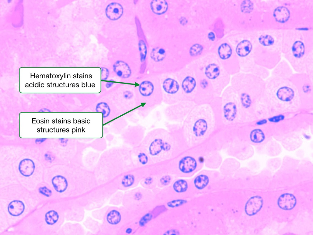
The H E staining method involves application of haematoxylin mixed with a metallic salt or mordant often followed by a rinse in a weak acid solution to remove excess staining differentiation followed by bluing in mildly alkaline water H E staining The most commonly used staining system is called H E Haemotoxylin and Eosin H E contains the two dyes haemotoxylin and eosin Eosin is an acidic dye it is negatively charged general formula for acidic dyes is Na dye It stains basic or acidophilic structures red or pink This is also sometimes termed eosinophilic
How Does H E Staining Work

How Does H E Staining Work
https://i.ytimg.com/vi/RDXtxa-mee4/maxresdefault.jpg

HE Staining Principle Procedure And Interpretation Haematoxylin
https://i.ytimg.com/vi/HUE8w5pGlWQ/maxresdefault.jpg

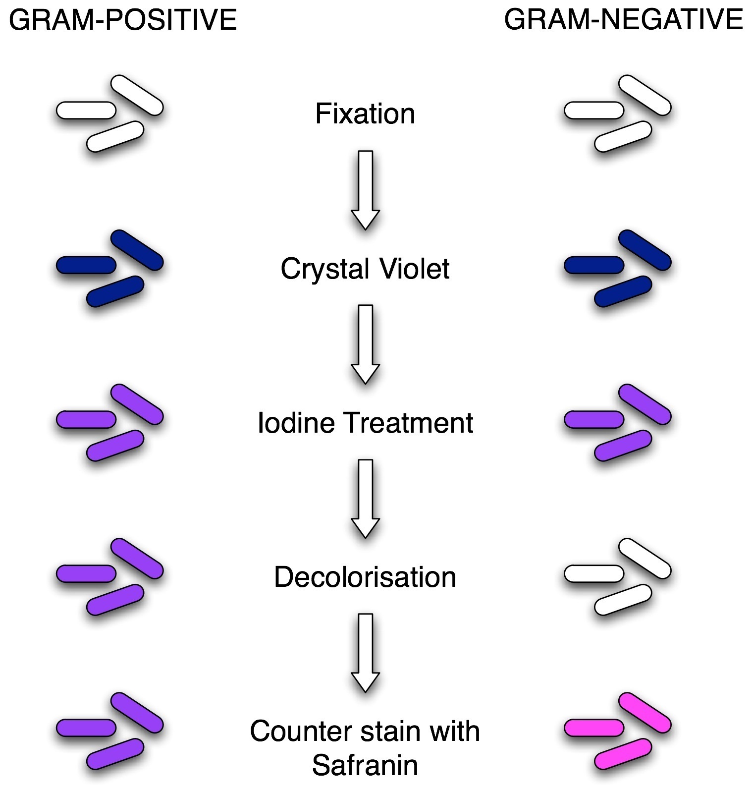
Gram Staining Diagram Diagram Quizlet
https://o.quizlet.com/KtrJRXdibNrl2h8piFdJfg_b.png
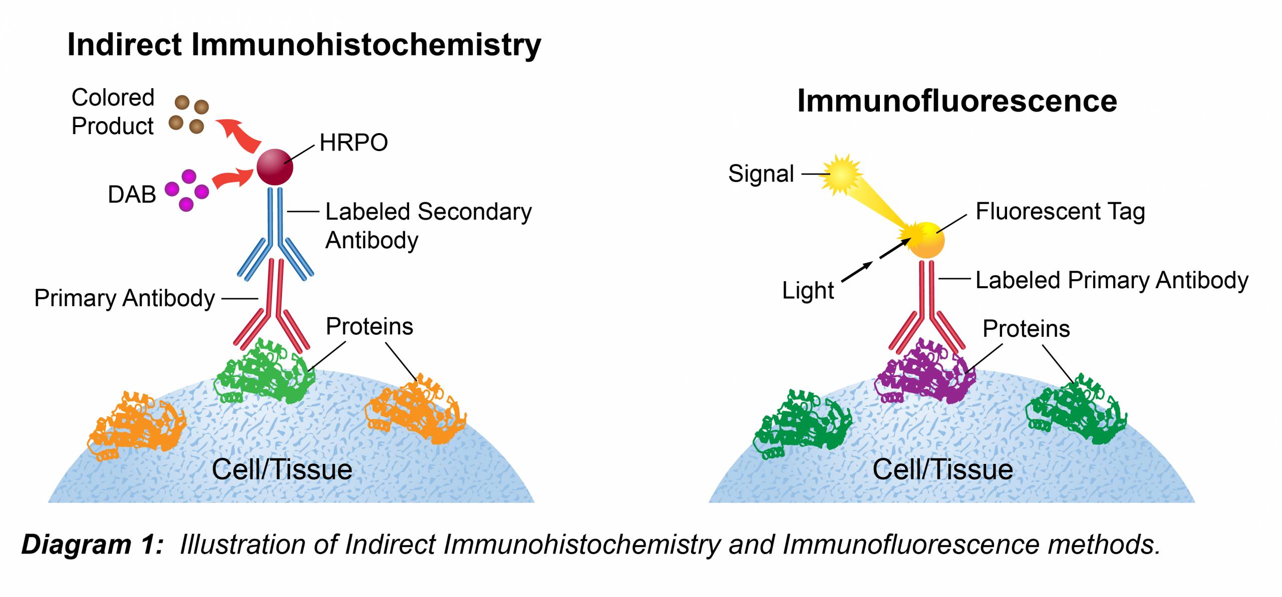
Hematoxylin generally without eosin is useful as a counterstain for many immunohistochemical or hybridization procedures that use colorimetric substrates such as alkaline phosphatase or peroxidase This protocol describes H E staining of H E Stain plays a pivotal role in biology histology pathology and cytology by revealing the intricate morphological details of tissues and lesions It operates on the principle of differential affinity between cell components and specific dyes
Hematoxylin and Eosin H E staining is a widely used histological staining technique in pathology and histology It is used to colourize tissues and cellular structures to observe and study them under a microscope Staining is a process by which a colour is imparted to sectioned tissue Specially manufactured dyes are used for this purpose In the following sections we will dive deeper into how hematoxylin and eosin function in tissue staining walk through the step by step staining protocol discuss its applications in histopathology and explore how to interpret H E stained tissue sections
More picture related to How Does H E Staining Work

Tbs Buffer Recipe Ihc Bryont Blog
https://www.leinco.com/wp-content/uploads/2020/04/immunohistochemistry-02-scaled.jpg

Cresyl Violet Stain Histology
https://s3.us-east-2.amazonaws.com/yalehistology/histological_features_of_cells/h_and_e_stained_sample.jpg

Gram Stain Lab Questions Riset
https://i.pinimg.com/originals/d0/a5/7f/d0a57f06343eee416ebaf70f3edfe353.jpg
For routine examination H E is the stain of choice It adds color to otherwise transparent tissue sections so that you can get a detailed view of the tissue structure under a microscope As its name suggests H E stain is a combination Hematoxylin and eosin H E are the principal stains applied for the demonstration of the nucleus and the cytoplasmic inclusions Harri s hematoxylin primary stain contains alum and alum acts as a mordant that stains the nucleus light blue which turns red in
[desc-10] [desc-11]

FIGURE Representative Images Of H E Staining In Colon And Liver
https://www.researchgate.net/publication/362917101/figure/fig3/AS:11431281080989559@1661453931896/FIGURE-Representative-images-of-H-E-staining-in-colon-and-liver-sections-We-use-green.png

Simple Stain Methylene Blue
https://universe84a.com/wp-content/uploads/2021/08/Methylene-Blue-Stain-Introduction-Principle-Composition-Preparation-Staining-Procedure-Result-Interpretation-and-Keynotes-1024x576.jpg

https://laboratorytests.org › hematoxylin-and-eosin-staining
Hematoxylin and Eosin H E staining is the most widely used staining technique in histopathology As its name suggests H E stain makes use of a combination of two dyes namely hematoxylin and eosin This combination deferentially stains various tissue elements and make them easy for observation

https://en.wikipedia.org › wiki › H&E_stain
The H E staining method involves application of haematoxylin mixed with a metallic salt or mordant often followed by a rinse in a weak acid solution to remove excess staining differentiation followed by bluing in mildly alkaline water

Why Gram Positive Bacteria Are Purple In Color After Using Safranin

FIGURE Representative Images Of H E Staining In Colon And Liver

Gram Staining Principle Procedure Results Microbe Online

Methylene Blue Stain Bacteria

Staining And Morphology Factors That Can Impact Accurate AI driven

A Beginner s Guide To Haematoxylin And Eosin Staining

A Beginner s Guide To Haematoxylin And Eosin Staining

Graphic Protocol For The Preparation And Fluorescent IHC Staining Of

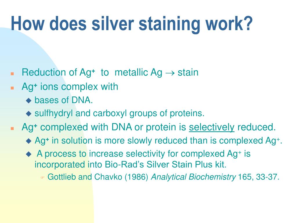
PPT Silver Staining PowerPoint Presentation Free Download ID 6685211

Types Of Staining Techniques Used In Microbiology Microbe Online
How Does H E Staining Work - Hematoxylin generally without eosin is useful as a counterstain for many immunohistochemical or hybridization procedures that use colorimetric substrates such as alkaline phosphatase or peroxidase This protocol describes H E staining of